Overview & Procedure
A perimetry test (visual field test) measures all areas of your eyesight, including your side, or peripheral, vision.
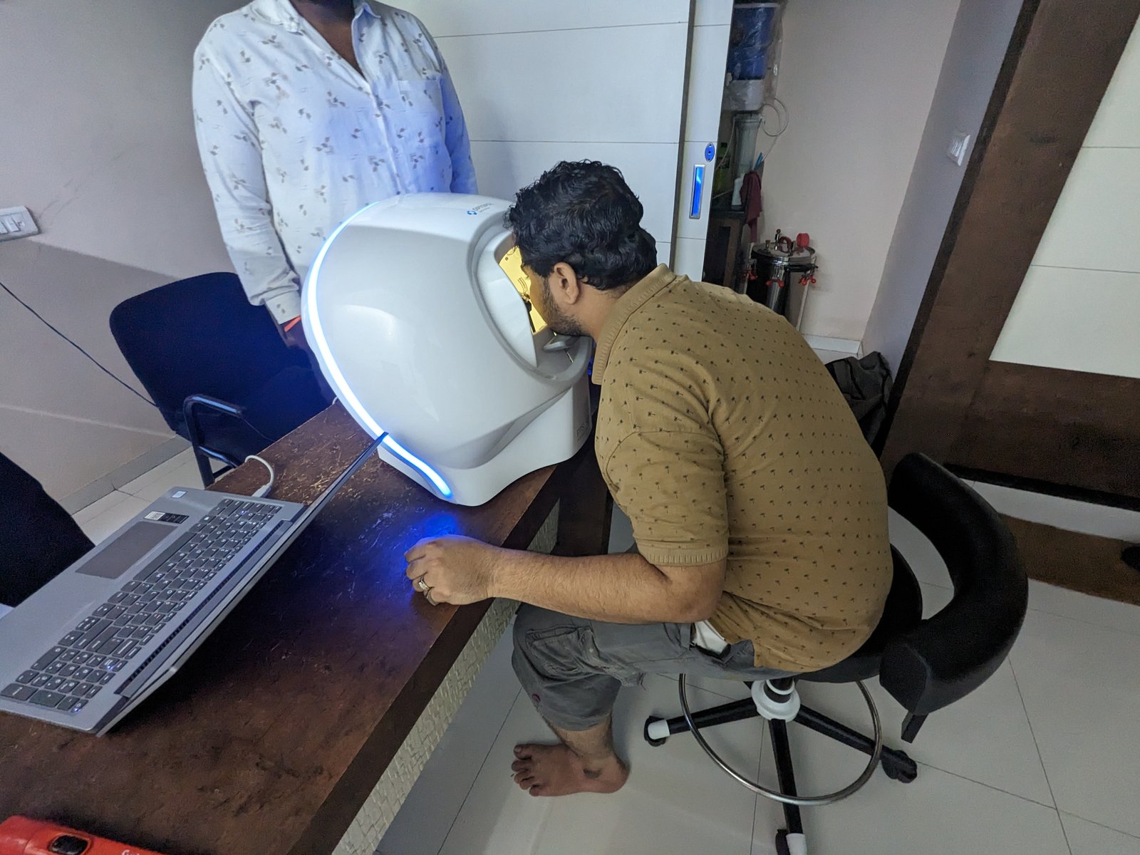
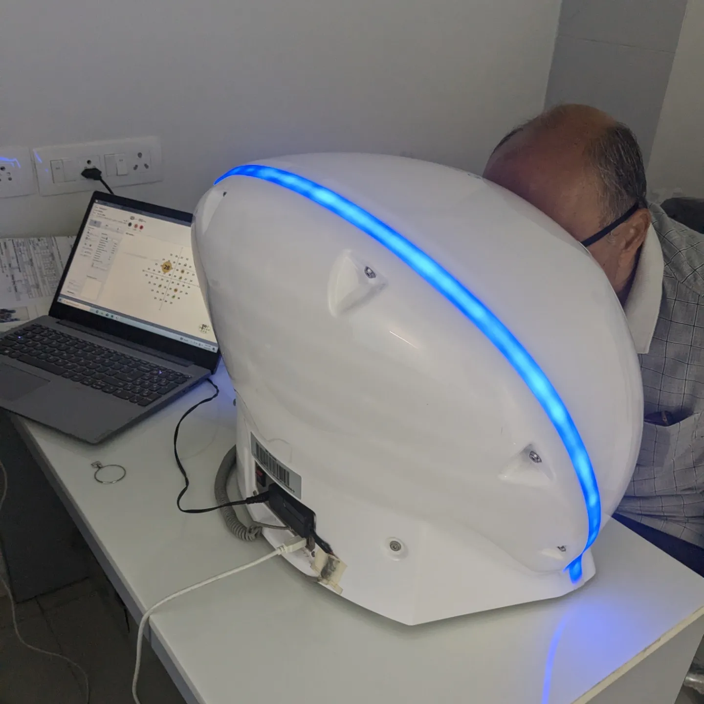
To do the test, you sit and look inside a bowl-shaped instrument called a perimeter. While you stare at the center of the bowl, lights flash. You press a button each time you see a flash. A computer records the spot of each flash and if you pressed the button when the light flashed in that spot.
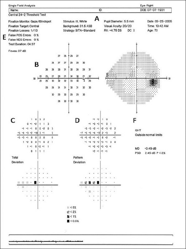
At the end of the test, a printout shows if there are areas of your vision where you did not see the flashes of light. These are areas of vision loss. Loss of peripheral vision is often an early sign of glaucoma.
Static vs Dynamic perimetry
Stimuli presented by perimeters can be static or kinetic.
- In kinetic perimetry, a stimulus is moved from a non-seeing (subthreshold) area to a seeing (suprathreshold) area, and the location where the object is first seen is recorded. The speed the stimulus is moved should be standardized, typically at 2-4 degrees per second.
- In static perimetry, stationary stimuli are presented at defined points in the visual field. Stimuli presented for longer durations of time may be seen better as a result of temporal summation of information, though limited additional benefit is derived beyond times over 1/10th of a second.
The Humphrey and Octopus perimeters use stimuli presented for 0.2 seconds and 0.1 seconds, respectively, maximizing temporal summation while minimizing patient attempts to redirect fixation towards the stimulus.


Normal field of vision
The normal eye can detect stimuli over a 120º range vertically and a nearly 160 degree range horizontally. From the point of fixation, stimuli can typically be detected 60º superiorly, 70º inferiorly, 60º nasally, and 100 degrees temporally, though the true extent of the visual field depends on several features of the stimulus (size, brightness, motion) as well as the background conditions. The field of vision is often depicted as a three dimensional hill, with the peak sensitivity to stimuli occurring at the point of fixation under photopic conditions, decreasing rapidly in the 10º around fixation, and then decreasing very gradually for locations further in the periphery. Nerve fibers pass through the sclera at the optic nerve head, typically 10-15º nasal to fixation. At this location, no photoreceptors are present, creating a normal absolute scotoma.
Visual field defects in glaucoma
- A nasal step defect obeying the horizontal meridian
- A temporal wedge defect
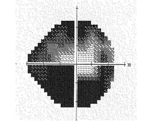
- The classic arcuate defect, which is a comma-shaped extension of the blind spot
- A paracentral defect 10-20° from the blind spot
- An arcuate defect with peripheral breakthrough
- Generalised constriction (tunnel vision)
- Temporal-sparing severe visual field loss
- Total loss of field.

