Retina Treatment
Introduction
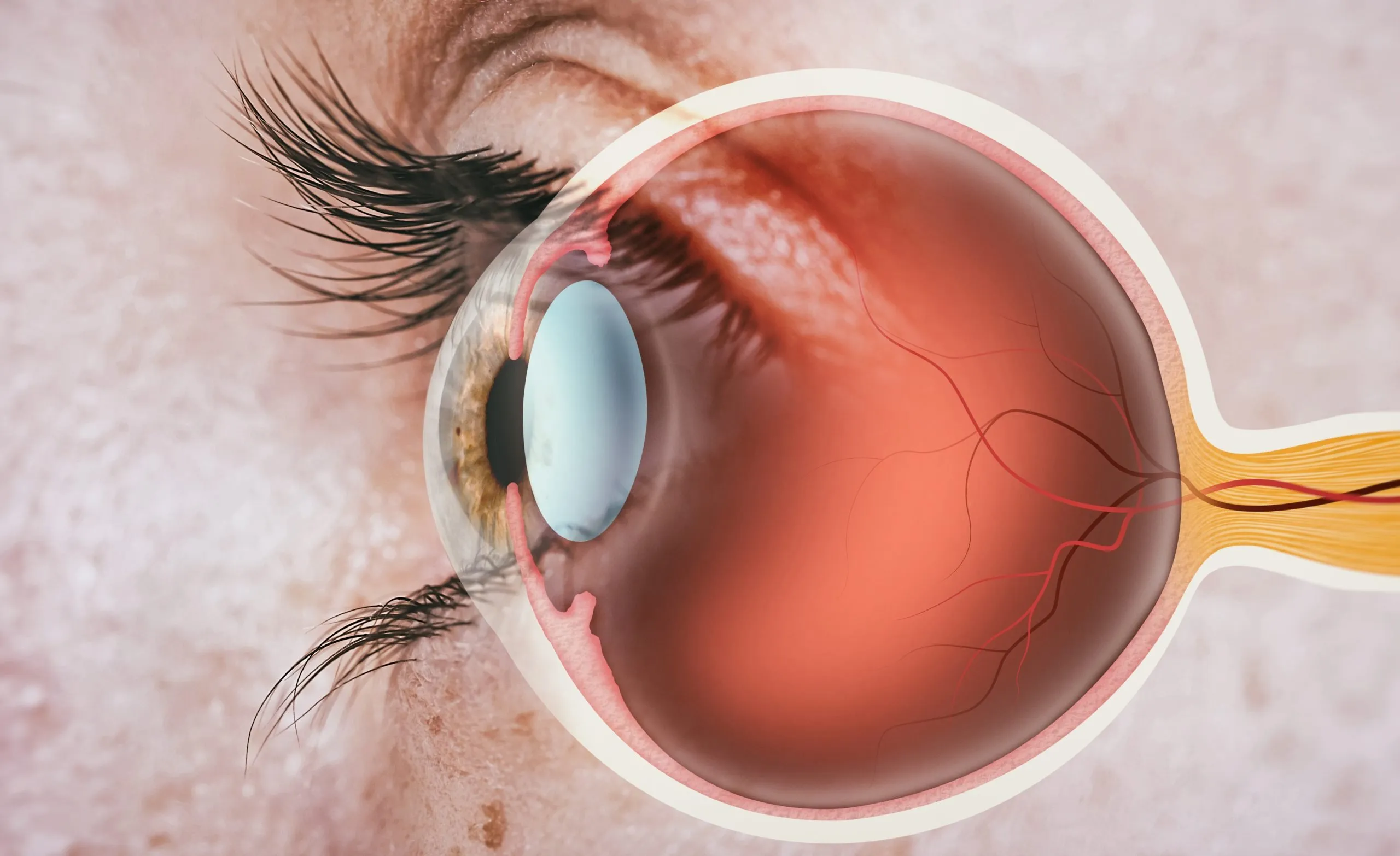
Retina treatment encompasses a range of medical interventions aimed at preserving or restoring vision compromised by retinal disorders. The retina, a delicate layer of tissue at the back of the eye, plays a crucial role in vision by converting light into neural signals that are sent to the brain. When the retina is damaged or affected by disease, it can result in vision loss or impairment. Fortunately, advancements in medical technology and treatment options offer hope to those suffering from retinal conditions.
Benefits and Considerations
Retina treatment can offer significant benefits, including preserving remaining vision, improving visual acuity, and enhancing quality of life. However, it's essential to consider potential risks and limitations associated with treatment options, as well as the need for ongoing monitoring and follow-up care.
How is Retina checkup done?
A retinal exam is not much different than any other eye exam. A retinal eye exam usually involves these steps:
- First, you’ll be asked about your medical history and any vision problems or symptoms you might be experiencing and what brought you here.
- Next, how clearly you can see (visual acuity) is measured. Your eye pressure is measured, during which you may receive drops that enlarge your pupils.
- Your doctor checks the health of your eyes and retinas, using several lights and performing different types of tests.
Retinal examination
A retinal examination — sometimes called ophthalmoscopy or funduscopy — allows your doctor to evaluate the back of your eye, including your retina, optic disk and the underlying layer of blood vessels that nourish the retina (choroid). Usually before your doctor can see these structures, your pupils must be dilated with eye drops that keep the pupil from getting smaller when your doctor shines light into the eye.
After administering eye drops and giving them time to work, which on rare occasion may burn a bit, your eye doctor may use one or more of these techniques to view the back of your eye :
- Direct examination. Your eye doctor uses an ophthalmoscope to shine a beam of light through your pupil to see the back of your eye.
- Indirect examination (indirect ophthalmoscopy). Your eye doctor examines the inside of the eye with the aid of a condensing lens and a bright light mounted on his or her forehead. This exam lets your eye doctor see the retina and other structures inside your eye in great detail and in three dimensions.
- Slit-lamp exam. In this exam your doctor shines the beam of a slit lamp through a special lens into your eyes. The slit lamp reveals a more-detailed view of the back of your eye.
The retinal examination usually takes less than 10 minutes. Your doctor will discuss the results with you following the exam.
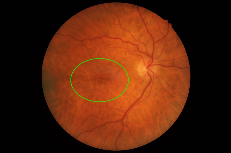
Age-related macular degeneration (AMD) :
This progressive condition affects the macula, the central part of the retina responsible for sharp, central vision. AMD can lead to blurred or distorted vision and, in advanced stages, central vision loss.
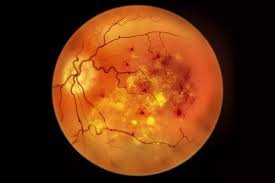
Diabetic retinopathy :
A complication of diabetes, diabetic retinopathy occurs when high blood sugar levels damage blood vessels in the retina. This can lead to vision problems and even blindness if left untreated.
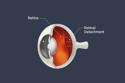
Retinal detachment :
This occurs when the retina pulls away from its normal position, leading to vision loss. It requires immediate medical attention to prevent permanent vision loss.
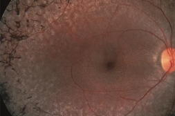
Retinitis pigmentosa :
A group of genetic disorders that cause progressive degeneration of the retina, resulting in night blindness and a gradual loss of peripheral vision.
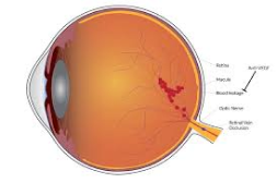
Macular edema :
Swelling of the macula, often associated with conditions such as diabetic retinopathy or AMD, can lead to distorted or blurred central vision.
Treatment Options :

Intravitreal injections :
Medications, such as anti-VEGF drugs or corticosteroids, can be injected into the vitreous gel of the eye to reduce inflammation, block abnormal blood vessel growth, or control swelling.
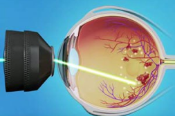
Laser therapy :
Laser treatment, such as photocoagulation or photodynamic therapy, can be used to seal leaking blood vessels, destroy abnormal tissue, or promote the regression of abnormal blood vessels.
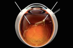
Vitrectomy :
In cases of severe retinal detachment or advanced proliferative diabetic retinopathy, a vitrectomy may be performed. This surgical procedure involves removing the vitreous gel and repairing or removing scar tissue to reattach the retina.
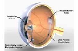
Retinal prostheses :
In some cases of advanced retinal degeneration, retinal implants or prostheses may be used to replace damaged photoreceptors and restore partial vision.

Gene therapy :
Emerging treatments, such as gene therapy, aim to address the underlying genetic causes of certain retinal disorders by introducing functional genes into retinal cells.
